Pelvic & Fertility Ultrasound Scan: Comprehensive Reproductive Health Assessment
Whether you’re planning a family, monitoring symptoms, or simply keeping track of your reproductive health, a Pelvic & Fertility Ultrasound Scan is one of the most effective ways to understand your body and make informed decisions. This safe, non-invasive scan provides detailed insights into your uterus, ovaries, and overall pelvic health.
At Ultramsono, based in Kensington Olympia, London, we offer private Pelvic & Fertility Ultrasound Scan services with fast access, same-day results, and professional care, helping you take proactive steps in managing your reproductive wellbeing.
What Is a Pelvic & Fertility Ultrasound Scan?
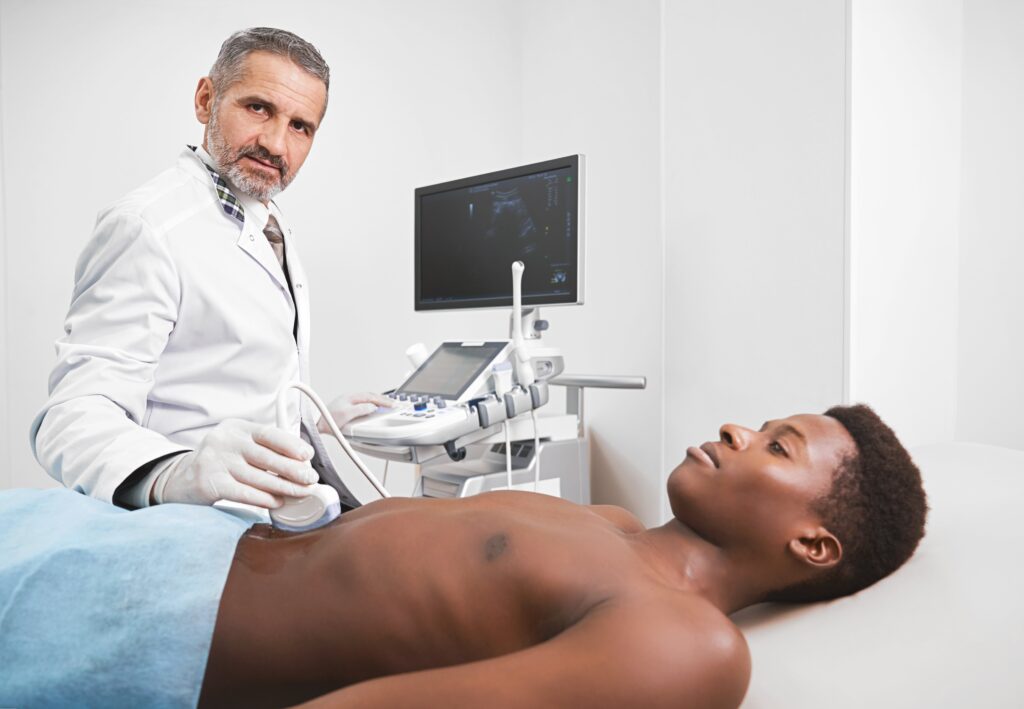
A Pelvic & Fertility Ultrasound Scan uses high-frequency sound waves to produce detailed images of the reproductive organs, including the uterus, endometrium (lining), ovaries, and surrounding pelvic structures. The procedure is painless, non-invasive, and free of radiation, making it safe for routine health checks, fertility monitoring, or early investigation of symptoms.
There are two main types of scans:
-
Transabdominal scan: performed over the lower abdomen; a full bladder improves visibility.
-
Transvaginal scan: provides a closer, more detailed view of the pelvic organs when clinically indicated.
Our skilled sonographers guide you through the scan, ensuring your comfort and understanding at every step.
How a Pelvic & Fertility Ultrasound Scan Can Help
For individuals trying to conceive or exploring fertility treatments, a Pelvic & Fertility Ultrasound Scan is crucial for evaluating factors affecting conception. This scan helps assess:
-
Ovarian function: including follicle count and ovulation monitoring
-
Uterine health: detecting fibroids, polyps, or structural abnormalities
-
Endometrial thickness: supporting implantation assessment
-
Polycystic ovaries (PCO/PCOS): evaluating hormonal and ovulatory balance
These findings provide your GP, gynecologist, or fertility specialist with essential information to create a personalized treatment or care plan.
When to Consider a Pelvic & Fertility Ultrasound Scan
You may benefit from a Pelvic & Fertility Ultrasound Scan if you experience:
-
Irregular, heavy, or painful periods
-
Persistent pelvic pain or bloating
-
Difficulty conceiving or fertility challenges
-
Family history of ovarian cysts or fibroids
-
Hormonal imbalance symptoms such as acne, hair growth, or weight changes
Even without symptoms, a routine Pelvic & Fertility Ultrasound Scan serves as a valuable baseline for reproductive health monitoring and early detection of potential issues.
Why Choose a Private Pelvic & Fertility Ultrasound Scan at Ultramsono
At Ultramsono, we provide:
✅ Quick appointments without long waiting lists
✅ Same-day, clear, and detailed reports
✅ A calm, discreet, and professional clinical environment
✅ Expert sonographers with years of experience in reproductive imaging
✅ Guidance and support for any necessary follow-up care
Every Pelvic & Fertility Ultrasound Scan is performed with sensitivity, professionalism, and patient comfort as a priority.
Empowering Your Reproductive Health
Understanding your reproductive health is empowering. A Pelvic & Fertility Ultrasound Scan can provide clarity, reassurance, and actionable insights. Whether you’re planning a family, managing symptoms, or simply gaining knowledge about your body, this scan ensures you are informed and supported throughout your health journey.
Book Your Private Pelvic & Fertility Ultrasound Scan in London
Take control of your reproductive health with a private Pelvic & Fertility Ultrasound Scan at Ultramsono. Appointments are available promptly, and a full report is provided on the same day.
📞 Call or WhatsApp: 07828 668209
📧 Email: info@ultramsono.co.uk
🌐 Visit: www.ultramsono.co.uk
Ultramsono – Medical Imaging by Ultram Ltd
📍 Kensington Olympia, London