Navigating the World of Ultrasound Imaging: A Comprehensive Guide to Safety and Efficacy
Ultrasound imaging, a versatile and widely used diagnostic technique, has revolutionized the medical field by providing real-time, non-invasive images of internal organs and structures. Its ability to produce clear, detailed images without the use of ionizing radiation has made it a preferred imaging modality for a wide range of medical applications, particularly in pregnancy.
Safety: A Foundational Principle
The safety of ultrasound imaging is a paramount concern, and extensive research has demonstrated its overall safety profile. Unlike ionizing radiation-based imaging techniques such as X-rays and CT scans, ultrasound does not utilize ionizing radiation, eliminating the risk of radiation-induced damage to cells and tissues.
High-Frequency Sound Waves: The Mechanism of Imaging
Ultrasound imaging harnesses the power of high-frequency sound waves, beyond the range of human hearing, to generate images of internal structures. These sound waves are directed into the body, where they interact with organs and tissues, causing echoes. These echoes are then collected and processed by ultrasound equipment, producing detailed images of the internal anatomy.
Non-Invasiveness and Painlessness: Enhancing Patient Comfort
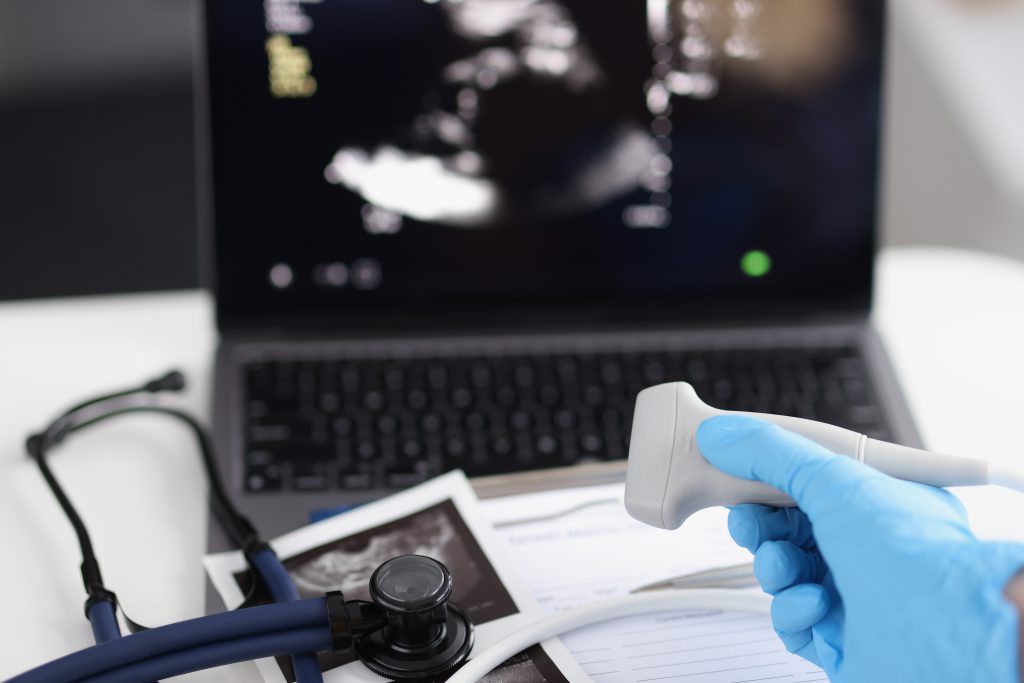
Ultrasound imaging stands as a non-invasive procedure, causing no pain or discomfort to patients. The transducer, the handheld device used to transmit and receive sound waves, is gently placed on the skin, allowing for the acquisition of images without the need for needles or injections.
Ultrasound in Pregnancy: A Safe and Effective Tool
Ultrasound has revolutionized prenatal care, providing invaluable insights into fetal development and enabling early detection of abnormalities. Numerous studies have confirmed the safety of ultrasound during pregnancy, with no evidence of harmful effects on the developing fetus or the mother.
Limiting Unnecessary Exposure: A Prudent Approach
While ultrasound is generally considered safe, it is prudent to minimize unnecessary exposure. Repeated ultrasound exams should only be performed when medically necessary, and the duration and intensity of each exam should be kept to a minimum.
Qualified Professionals: Ensuring Expertise and Safety
Ultrasound imaging should only be performed by qualified healthcare professionals who have received specialized training in ultrasound techniques. Proper training ensures that ultrasound equipment is used appropriately and that patients receive accurate diagnoses while minimizing potential risks.
ALARA: A Guiding Principle for Minimizing Exposure
The ALARA (As Low As Reasonably Achievable) principle is a cornerstone of medical imaging, emphasizing the minimization of patient radiation exposure while maintaining high-quality diagnostic images. Although ultrasound does not utilize ionizing radiation, the ALARA principle can still be applied to minimize exposure to sound waves.
Strategies for Implementing ALARA in Ultrasound Imaging
Several strategies can be employed to implement the ALARA principle in ultrasound imaging:
- Ultrasound Use Only When Medically Necessary: Physicians should only order ultrasound exams when they are clinically indicated, avoiding unnecessary or repeat exams.
- Optimizing Ultrasound Settings: Ultrasound technologists should employ the lowest possible intensity and duration settings that still produce accurate diagnostic images.
- Minimizing Exam Duration: Ultrasound exams should be kept as brief as possible to reduce patient exposure to sound waves.
- Ultrasound Guidance for Procedures: Ultrasound guidance can be used during medical procedures to precisely target the procedure site, minimizing the need for additional imaging exams.
- Regular Equipment Maintenance: Regular maintenance and calibration of ultrasound equipment ensure optimal performance and accurate diagnostic images.
ISUOG Guidance on Doppler Ultrasound
The International Society of Ultrasound in Obstetrics and Gynecology (ISUOG) has issued guidelines on the safe utilization of Doppler ultrasound for fetal ultrasound assessments during the first 13+6 weeks of gestation.
Doppler Use During the Embryonic Period
The embryonic period spans from conception to 9 weeks after the last menstrual period. During this phase, rapid cell division and organ development occur. The fetal-placental circulation is not yet established. ISUOG recommends avoiding the routine use of Doppler ultrasound modalities, such as spectral Doppler, colour flow imaging, and power imaging, during the embryonic period. If Doppler use is clinically necessary, exposure time should be minimized.
Doppler Use During the Fetal Period
The fetal period begins at approximately 11 weeks after the last menstrual period, when the crown-rump length reaches 45 mm. During this time, organ development is complete, and the fetal-placental circulation is established. ISUOG guidelines indicate that certain clinical indications, such as trisomy and cardiac anomaly screening, may warrant the use of Doppler ultrasound modalities from 11+0 to 13+6 weeks. When performing Doppler ultrasound, the displayed thermal index (TI) must not exceed 1.0, and exposure time should be kept to a minimum, typically no more than 5-10 minutes.
ULTRAM’s Commitment to ALARA
At ULTRAM, we are committed to upholding the ALARA principle in all ultrasound imaging procedures. Our team of experienced and certified ultrasound technologists adheres to strict protocols to minimize patient exposure to sound waves while maintaining the highest quality diagnostic images. We employ state-of-the-art ultrasound equipment that is regularly maintained and calibrated to ensure optimal performance. Our dedication to ALARA ensures that our patients receive the safest and most effective ultrasound imaging experience possible.

